The last post covering the unknowns described my methods for obtaining and plating out my potential unknowns. Of the plated group, only the non surface sterilized mites, non sterilized thrips, and the one week old bean leaves yielded any fungi.
Non-sterilized thrips
 |
| A sub-culture of the fungus I obtained from thrips. |
On my non-sterilized thrips, I managed to get colonies of the same looking fungus coming from each thrips. I took a sterile transfer from the initial plate and started a pure culture. Upon examining the sample under the microscope from a riddel mount, I realized that this fungus was in fact
Penicillium, a fungus that we cannot use for this course. So, on to the other two for a better shot at a useable unknown.
 |
| Under the microscope: Penicillium the thrips fungus. |
Non-sterilized mites
 |
| A subculture from the fungus I obtained from spider mites. The center looks like a contamination by Penicillium . |
On the plate of 10 spider mites, I had a same morphology fungus growing out of each of the spider mites, so I took a subculture from one of them to start a pure culture. Apparently I used poor technique because it appeared that
Penicillium had made it into my plate as well. For future work, I used only the dark looking fungus. I had a bit of trouble identifying this fungus. When I attempted to key it out, I would come to multiple conclusions, dead ends, or couplets that I didn't really have enough evidence to decide. With the help of Dr. Shaw, we have decided to call this fungus
Drechslera, but I am going to sequence this soon to confirm this identification. I based my decision off of the shape of the conidia; they were cylindrical and rounded at the end, multi-spored, generally with straight walls dividing the cells. The conidia did not appear to grow in clusters either. Over time the conidia became darker, but initially they appeared fairly light.
 |
| Conidia on the conidiophore of Drechslera. |
When comparing the above image that I took, to the picture from the Barnett book, you can see they generally look similar in their formation.
Fed upon leaf material
I did not get very much material growing from the sterilized fed upon leaf material, but I did get one fungus that I was able to sub-culture to water agar. For almost two months I could not get any conidia to be produced, so I transfered a plug to 1/2 PDA, allowed it to grow in a L:D 14:10 for a week, then placed it in continuous light for 48 hours, then total dark for 48 hours. I'm not sure if it was the light schedule or the nutrients provided by PDA, but I was able to get conidia to form eventually.
 |
| The subculture I took from the initial plate with used leaf material. |
 |
| Conidia that I was eventually able to produce from this fungus. |
Once I was able to produce conidia, it was pretty apparent that this was Nigrospora. The short, urn-shaped conidiophore, and the dense, shiny black conidia and very obvious in this fungus. This is pretty textbook looking, so I am very confident that this is Nigrospora.
 |
| Nigrospora in the textbook. This is exactly what my fungus looks like. |
Additional sources for fungus
At this point I had only identified two fungi, but needed three. I initially wanted to sample various plants around the lab to see what endophytes they had, but time was not something that I had a lot of, so I settled for sampling rather random things. I collected some water from my fish tank, frass from caterpillars, fungi growing on some pinned insects, open plates left in the lab etc. While each of these gathered quite a few colonies of fungi, most of them were group I fungi, or so delicate that I was never really able to get a good look at the conidia on the conidophore. The one plate that I was able to get my final fungus came from a mushroom that I collected in my back yard...
Mushroom collected fungi
After we had a bloom of mushrooms around town, I decided to take one from my back yard into the lab to plate. I simply plucked the mushroom from the ground, placed it in a sterile bag, and then at the lab I used a sterile blade to excise a 1 cm cube from the gills.
 |
| The mushroom I selected from the yard. |
 |
| A close up of the gills. There is a lot of dirt and junk under here that should lead to some various fungal colonies when plated. |
The excised piece was smeared on a couple of 1/2 PDA plates, and then the piece was placed in its own plate. The plates were allowed to grow for a few weeks, and I got several different types of colonies.
 |
| One of the plates from mushroom with various colonies growing. |
I selected several colonies that were interesting looking to me to isolate for further investigation. Most of them were either the common fungi or too difficult to identify. One, however, maintained its composition when viewed with tape mounts, and I was able to identify the fungus.
 |
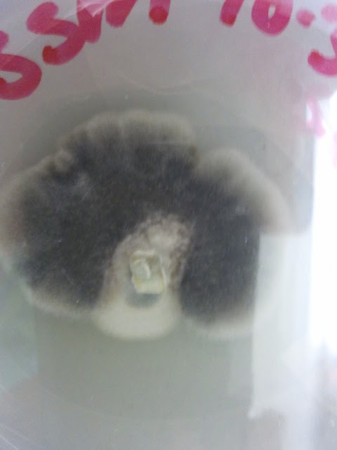
| A view through the plate of the third fungus I identified. |
I identified my final unknown as Monocillium. This is saprophytic in soils, so it is not very surprising that I would find it on a mushroom. The conidiophore is swollen, followed by a chain of single celled hyaline conidia.
 |
| Monocillium under the microscope. |
In this picture you can see the swollen conidiophore and chains of conidia. This looks very much like the image in the book.
 |
| Monocillium |
And that is it for the unknowns- Nigrospora, Drechslera, and Monocillium! It was quite the learning experience going through all the fungi that I grew trying to find some that would work for the class, but it was well worth it to help me in my future work. Thank you Dr. Shaw and Dr. Ebbole for your help this semester!


No comments:
Post a Comment